The Brain on Isometrics

Isometrics are often described as a muscular training method. That description is accurate, but incomplete. Before a muscle can produce isometric force, the brain must create, distribute, regulate, and continually refine the neural command responsible for it.
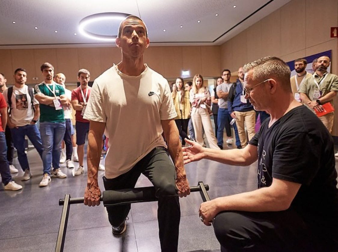
This matters because the training industry tends to interpret isometrics through what can be seen. The body appears quiet. There is no dramatic change in position, no visible acceleration, and no obvious athletic display. The exercise can therefore look neurologically simple.
The research suggests almost the opposite.
During an isometric contraction, the nervous system must recruit motor units, coordinate agonist muscles, integrate sensory information, regulate the magnitude and direction of force, correct fluctuations, suppress unwanted muscle activity, and—during bilateral tasks—manage communication between both cerebral hemispheres. What appears simple from outside may require sophisticated neurological organization inside.
The muscle produces the force. The brain organizes it.
Force Is a Neural Output
We commonly talk as though muscles independently produce force. A quadriceps contracts. A calf generates tension. A hand squeezes. Yet voluntary force does not originate in the muscle. It emerges from a motor command travelling through the nervous system and being expressed through motor neurons, motor units, muscle fibres, tendons, and the surrounding mechanical system.
The motor unit—the motor neuron and the muscle fibres it innervates—is the final common pathway through which the nervous system controls muscular force. However, the brain does not independently operate thousands of motor units like individual switches. Motor neurons receive combinations of shared and independent synaptic inputs that influence which motor units are recruited, how frequently they discharge, and how their outputs combine.
Del Vecchio and colleagues examined this organization during submaximal isometric contractions involving synergistic muscles of the hand and quadriceps. They identified multiple motor-unit modes representing different sources of common neural input. In the quadriceps, many motor units followed a muscle-specific module, others received shared input, and some aligned more closely with the neural module associated with the neighbouring agonist muscle.
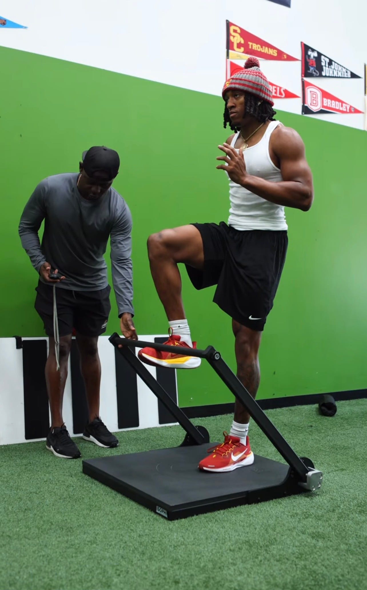
This is not merely an interesting neurological detail. It suggests that the nervous system can vary how force is distributed between agonist muscles even when the external isometric task appears unchanged. Independent and shared motor-unit modes may give the central nervous system greater precision in coordinating muscular output, directing force through the tendon, and regulating internal joint stresses.
An isometric knee extension is therefore not one fixed neurological event. The position may be identical, but the motor solution can change with force intensity, effort direction, fatigue, sensory feedback, previous injury, and the number of muscles required to complete the task.
Greater Isometric Force Requires Greater Cortical Activity
The neurological demand also appears to scale with isometric intensity.
Abeln and colleagues recorded brain and muscle activity while participants performed unilateral isometric leg extensions at graded intensities. As isometric force increased, activity within the vastus medialis and vastus lateralis increased, as expected. More importantly, cortical current density within the primary motor cortex also increased with isometric intensity. The same intensity-dependent increase was not observed in the premotor cortex, primary somatosensory cortex, or somatosensory association cortex.
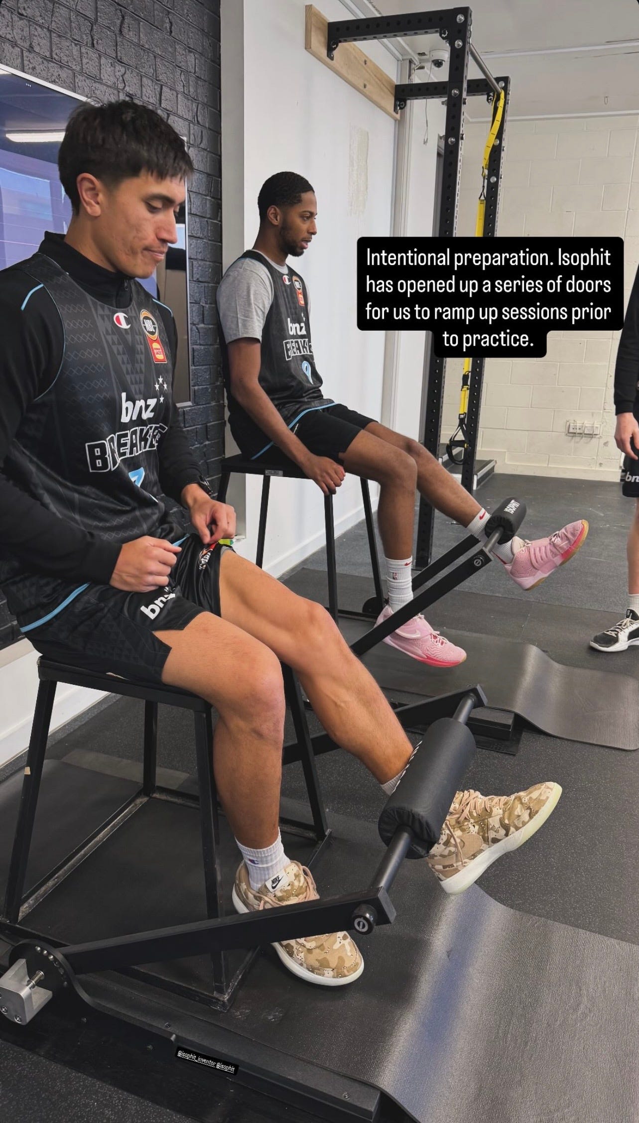
The study was small, and it measured acute activity rather than long-term adaptation. It cannot tell us that harder isometrics automatically create better neurological outcomes. It does, however, demonstrate an important principle: two isometric contractions performed in the same position but at different intensities are not simply lighter and heavier versions of the same muscular task.
They create different cortical demands.
This should change how coaches and clinicians prescribe isometric training. Twenty per cent of maximal voluntary contraction may be appropriate for motor learning, early rehabilitation, active recovery, or cardiovascular applications. Eighty per cent may create a substantially different demand for motor-unit recruitment and cortical drive. The joint position alone does not define the stimulus. Intensity changes what the nervous system must accomplish.
Bilateral Isometrics Make the Brain Negotiate
The neurological complexity becomes even more apparent when both sides of the body produce isometric force simultaneously.
Long and colleagues studied unilateral and bilateral isometric index-finger contractions at 10%, 40%, and 70% of maximal voluntary contraction. During stronger bilateral contractions, communication between the motor cortices changed. Alpha-band coherence decreased while transcallosal inhibition increased at 40% and 70%, but not at 10%, of maximal force. Participants who demonstrated this relationship were also better able to maintain steadier bilateral isometric contractions.
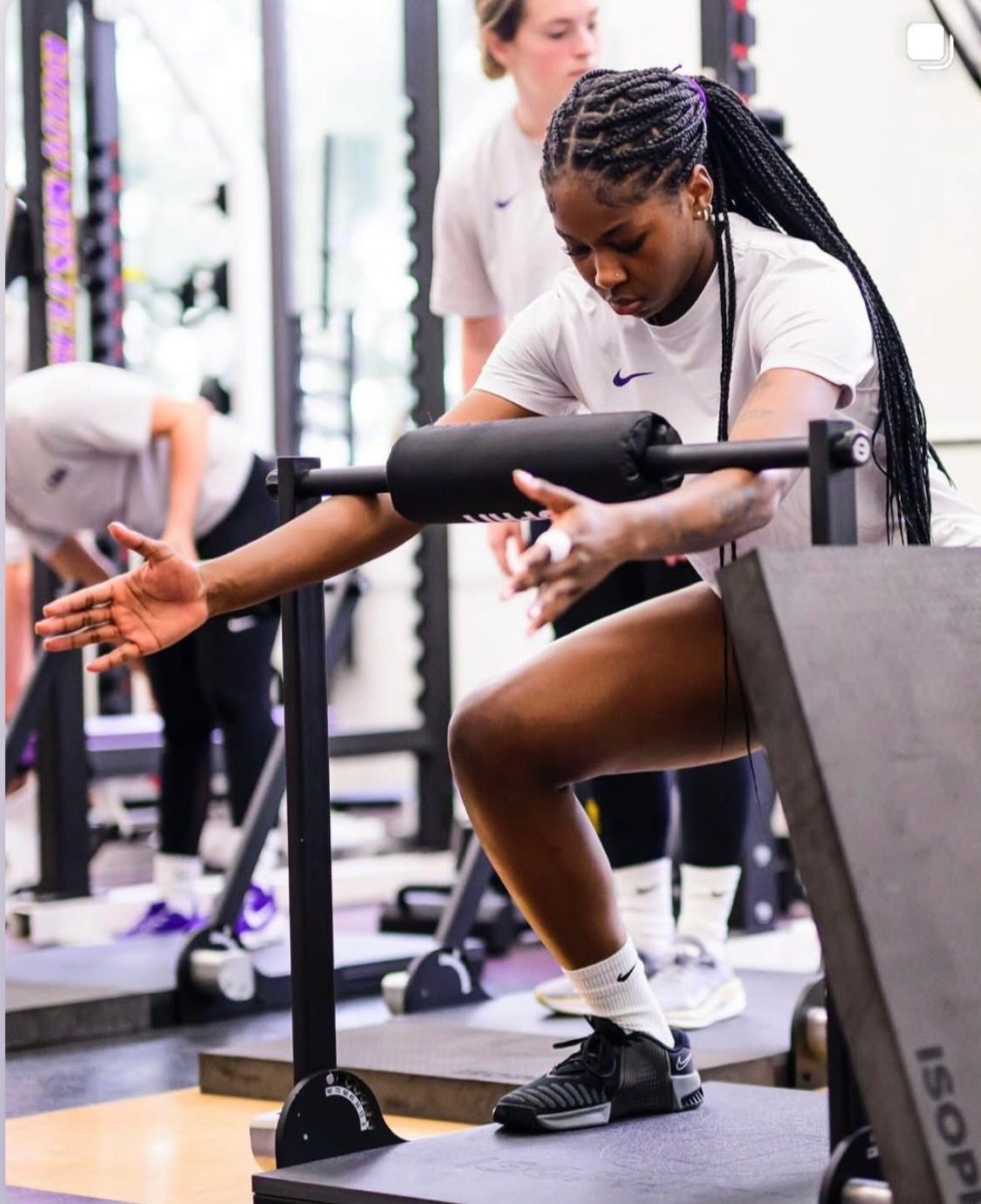
The researchers proposed that these cortical interactions may help suppress neural cross-talk, reduce task-irrelevant activity, prevent unwanted muscular activation, and improve the steadiness of strong bilateral force.
In other words, the two hemispheres are not merely sending parallel commands to the two hands. They are cooperating, inhibiting, correcting, and managing interference. Strong bilateral isometric force requires the brain to coordinate both sides while preventing one side’s neural activity from disrupting the other.
This makes force steadiness more than a muscular quality. It becomes evidence of neurological regulation.
Strength Is Not Enough
Traditional strength testing asks how much force a person can produce. That is valuable, but it captures only the output. It does not reveal how efficiently the nervous system organized that output, how much unwanted activity occurred, how stable the force remained, or whether the same force could be reproduced across repeated contractions.
Two athletes can produce the same peak isometric force and possess very different neurological capabilities. One may reach the target quickly, regulate it precisely, sustain it efficiently, and reproduce it under fatigue. The other may reach the same peak through greater co-contraction, larger force fluctuations, compensatory muscle activity, or an inconsistent motor strategy.
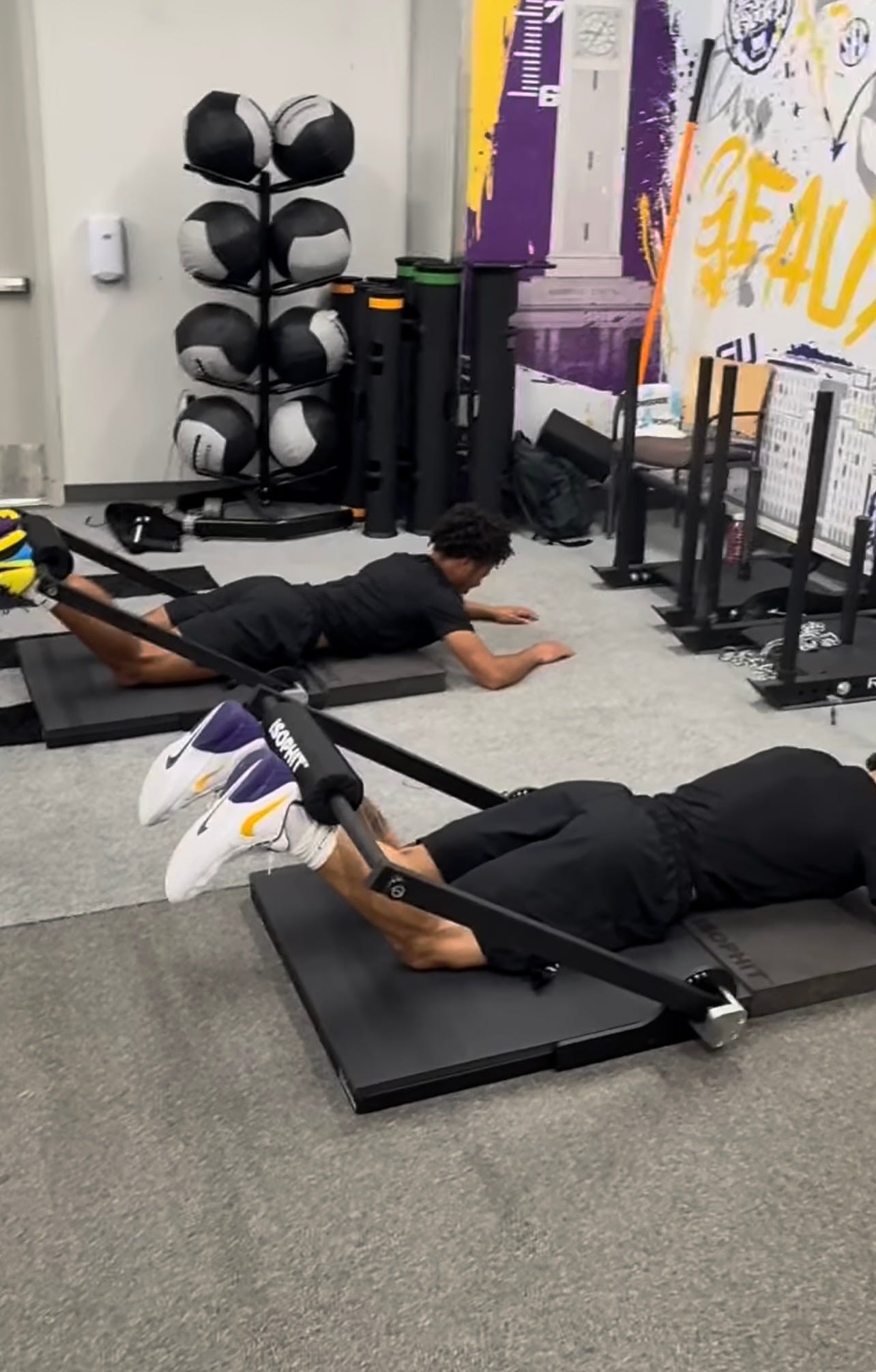
Peak isometric force matters. So do rate of isometric force development, force steadiness, time to target, bilateral symmetry, repeated-contraction consistency, and Total Force Over Time. Together, these measures begin to reveal not merely whether force was produced, but how well the nervous system governed it.
That distinction has implications for athletic performance, rehabilitation, neurological recovery, and aging. Human movement does not depend only on having strong muscles. It depends on the ability of the nervous system to produce, distribute, regulate, tolerate, and transmit isometric force before that force can be expressed through movement.
Isometrics are not an absence of neurological activity. They are an opportunity to observe force before movement conceals the system responsible for creating it.
At Isophit, we help the world’s strongest, fastest, and most dominant athletes—and everyday people—win more, hurt less, and age stronger.
Learn more at www.isophit.com.
Knowledge only creates value when it is shared. If this article changed how you think about the topic, consider sharing it with a coach, athlete, clinician, colleague, friend, or family member.